Woman Anatomy Of Chest Wall - Anatomy of chest wall and thoracic cavity medical images ... : Primary tumors originate in the bone or muscle of the chest wall.
Woman Anatomy Of Chest Wall - Anatomy of chest wall and thoracic cavity medical images ... : Primary tumors originate in the bone or muscle of the chest wall.. The chest wall is comprised of skin, fat, muscles, and the thoracic skeleton. The margin between the corpus and manubrium sterni is called the angulus sterni (ludovici or sternal angle) which is visible… The most common malignant chest wall tumors are sarcomas. The remaining part is made up of fatty tissue. Chest wall pain is caused by problems affecting the muscles, bones and/or nerves of the chest wall.
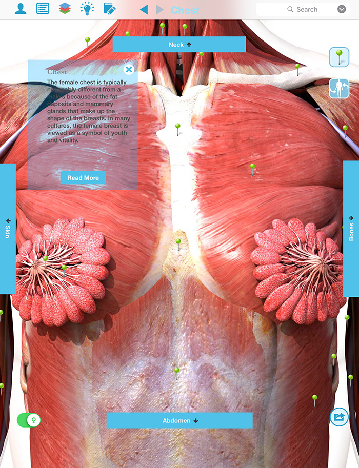
Chest muscles anatomy woman anatomy drawing diagram.medial half of anterior surface of clavicle. In humans, breast tissue begins to enlarge at puberty. The thoracic contents are bounded by the chest wall, providing both the shape of the thorax and protection for the intrathoracic contents ().the skin, subcutaneous tissues, and muscles that surround the rib cage and shoulder girdle appear radiographically indistinguishable from each other, whereas on ct the skin, fat, and muscles are recognized by their difference in attenuation. Skandalakis chest wall embryogenesis the muscles of the chest develop from the somites found in the mesoderm. The chest wall is a complex anatomic structure composed of muscles, bones, joints, and soft tissues that make up the area of the body between the neck and the abdomen.
Chest Pictures Of Anatomy from www.sciencealert.com Thoracic wall and breast illustrations : The chest wall functions as a protective cage around the vital organs of the body, and significant disruption of its structure can have dire respiratory and circulatory consequences. It provides protection to vital organs (eg, heart and major vessels, lungs, liver) and provides stability for movement. Applied anatomy of the chest wall and mediastinum petros mirilas michael e. The exam is done while lying down, not standing up. Thoracic wall dissection anatomy description: The most common benign tumors are osteochondromas and chrondromas. Chest wall pain is caused by problems affecting the muscles, bones and/or nerves of the chest wall.
The epidermis is the outermost layer that provides a protective, waterproof seal over the body.
Chest wall pain is caused by problems affecting the muscles, bones and/or nerves of the chest wall. In humans, breast tissue begins to enlarge at puberty. The right diaphragm should be visible all the way to the anterior chest wall (red arrow). Skandalakis chest wall embryogenesis the muscles of the chest develop from the somites found in the mesoderm. It provides protection to vital organs (eg, heart and major vessels, lungs, liver) and provides stability for movement. The exam is done while lying down, not standing up. Find the perfect female chest anatomy stock photo. Those patients either have a concave, hollow chest, in which the chest slopes towards the middle, or they may have an outward curved chest wall, or bird like chest, in which the middle part of the chest is the highest part like the women in the video still shown below. The anterior muscles of the trunk (torso) are associated with the front of the body lying below the pectoral muscles, the intercostal muscles form the chest wall and play a key role in respiration. Pathologic processes that may involve the chest wall include congenital and developmental anomalies, trauma, inflammatory and infectious diseases, and soft tissue and bone tumors. Because total chest wall volume increased by 4.46 l at t 3, assuming an average density equal to 1, the 6.5 kg of weight gained was therefore mainly located in the trunk (~69%), mostly in the abdomen (~65%), with 31% in the extremities. The thoracic contents are bounded by the chest wall, providing both the shape of the thorax and protection for the intrathoracic contents ().the skin, subcutaneous tissues, and muscles that surround the rib cage and shoulder girdle appear radiographically indistinguishable from each other, whereas on ct the skin, fat, and muscles are recognized by their difference in attenuation. The medical name for breast is mammary gland.
A woman's chest — like the rest of her body — is covered with skin that has two layers. 21.1) certain ribs and intercostal spaces can be located by palpating these structures. The remaining part is made up of fatty tissue. The chest wall is comprised of skin, fat, muscles, and the thoracic skeleton. Find the perfect female chest anatomy stock photo.
Vintage 1950's Frohse Chest & Abdomen Viscera Human ... from cdn.shopify.com The chest wall functions as a protective cage around the vital organs of the body, and significant disruption of its structure can have dire respiratory and circulatory consequences. Diagnosis of chest wall pain. The skeleton of the thoracic wall is formed by the twelve thoracic vertebra posteriorly, the sternum anteriorly and, on each side, by the twelve ribs and the respective costal cartilage. The major muscle in the chest is the pectoralis major. Applied anatomy of the chest wall and mediastinum petros mirilas michael e. Chest muscles anatomy woman : Primary tumors originate in the bone or muscle of the chest wall. The left diaphragm can only be seen to a point where it borders the heart (blue arrow).
In combination, these muscles play a highly important role in terms of shoulder and hand movements.
It is made up of the manubrium superiorly, the body and the xiphisternum (figure 1). A woman's chest — like the rest of her body — is covered with skin that has two layers. Chest wall pain is caused by problems affecting the muscles, bones and/or nerves of the chest wall. Chest muscles anatomy woman : Doctors diagnose chest wall pain in at least 25% of patients who come to the emergency room for chest pain. This is because when lying down the breast tissue spreads evenly over the chest wall and is as thin as possible, making it much easier to feel all the breast tissue. The most common malignant chest wall tumors are sarcomas. Huge collection, amazing choice, 100+ million high quality, affordable rf and rm images. 21.1) certain ribs and intercostal spaces can be located by palpating these structures. About the 6th week, the somites differentiate into the sclerotomes and the dermatomyotomes. The major muscle in the chest is the pectoralis major. The chest wall is a complex anatomic structure composed of muscles, bones, joints, and soft tissues that make up the area of the body between the neck and the abdomen. Use the finger pads of the 3 middle fingers on your left hand to feel for lumps in the right breast.
The spaces between the ribs are filled by the intercostal musculature, which consists of three layers. The most common malignant chest wall tumors are sarcomas. 21.1) certain ribs and intercostal spaces can be located by palpating these structures. Related posts of anatomy of the chest area anatomy of penis. Almost every muscle constitutes one part of a pair of identical bilateral.
Trunk Definition Anatomy from thumbor.kenhub.com Anatomy of the female breast (lateral view) they are supplied by several arteries of the thoracic wall, namely branches of the internal thoracic, axillary, lateral thoracic, thoracoacromial, and posterior intercostal arteries. The margin between the corpus and manubrium sterni is called the angulus sterni (ludovici or sternal angle) which is visible… The past several decades have seen a marked improvement in the management and reconstruction of complex chest wall defects. The epidermis is the outermost layer that provides a protective, waterproof seal over the body. Chest muscles anatomy woman anatomy drawing diagram.medial half of anterior surface of clavicle. This is because when lying down the breast tissue spreads evenly over the chest wall and is as thin as possible, making it much easier to feel all the breast tissue. Pectoralis major on women : The major muscle in the chest is the pectoralis major.
Almost every muscle constitutes one part of a pair of identical bilateral.
The spaces between the ribs are filled by the intercostal musculature, which consists of three layers. Actually we see the interface between the air in the lungs and the soft tissue structures in the abdomen. Thoracic wall and breast illustrations : Chest muscles anatomy woman : Two of the most common abnormalities of the chest wall are pectus excavatum and pectus carinatum, with the former being the most common congenital deformity of the sternum and the most common sternal deformity requiring surgery. The most common benign tumors are osteochondromas and chrondromas. Primary tumors originate in the bone or muscle of the chest wall. The palpable midline sternum is variable in size and shape; The skeleton of the thoracic wall is formed by the twelve thoracic vertebra posteriorly, the sternum anteriorly and, on each side, by the twelve ribs and the respective costal cartilage. Each breast consists of tissue overlying the chest wall muscles (the pectoral muscles). Applied anatomy of the chest wall and mediastinum petros mirilas michael e. In combination, these muscles play a highly important role in terms of shoulder and hand movements. It provides protection to vital organs (eg, heart and major vessels, lungs, liver) and provides stability for movement.
The left diaphragm can only be seen to a point where it borders the heart (blue arrow) anatomy of chest wall. The medical name for breast is mammary gland.